Detailed ultrasound imaging to assess the structure and function of your heart.
Expert cardiology care with a personal approach.
A transthoracic echocardiogram is a non-invasive ultrasound test used to assess the structure and function of the heart.
It provides detailed images of the heart’s chambers, valves and blood flow, helping diagnose a wide range of cardiac conditions including valve disease, heart failure and structural abnormalities.
This test is commonly used to investigate symptoms, monitor known heart conditions, and provide important information about how your heart is working.
During your consultation, your cardiologist will:
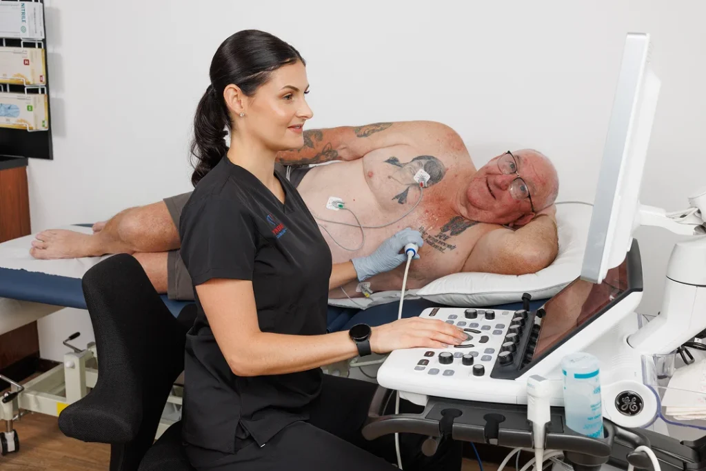
A trained cardiac technician performs the scan
A small probe is placed on the chest to capture ultrasound images
Images are taken from different angles to assess heart chambers, valves and blood flow
No radiation or injections are required
The test typically takes 30–45 minutes.
Your cardiologist will review the results and provide a report to your referring doctor.
You may be referred for an echocardiogram if you have:
Shortness of breath
Heart murmurs
Chest pain
Known or suspected heart conditions
Dr Jodi Harker is a highly experienced cardiologist providing comprehensive cardiac care across the Gold Coast.
With expertise in both diagnostic and preventative cardiology, Dr Harker focuses on delivering clear, patient-centred care tailored to each individual.
Her approach emphasises accurate diagnosis, clear communication and long-term heart health management.
Please bring:
This helps ensure your cardiologist has all the information needed to provide accurate assessment and advice.
If you have been referred for a cardiology consultation, our team is here to provide clear, structured and professional care.
An echocardiogram, or “echo,” is a type of ultrasound used to assess the structure and function of your heart. It creates real-time images that show how your heart muscle and valves are working, and how blood flows through the chambers.
The most common type is a transthoracic echocardiogram, where a small ultrasound probe is placed on the chest wall. This may also be called a resting echocardiogram, TTE, or cardiac ultrasound.
Echocardiograms are different from electrocardiograms (ECGs), which measure the electrical activity of your heart rather than its structure.
Other common types of echocardiograms include:
Exercise stress echocardiogram – performed before and after physical exercise
Dobutamine stress echocardiogram – uses medication to simulate exercise
Transoesophageal echocardiogram (TOE) – uses a probe inserted into the oesophagus under sedation to obtain detailed images.
An echocardiogram is a common and important test used to evaluate your heart’s structure and function. It helps your doctor better understand how your heart is working and can assist in diagnosing or monitoring a range of conditions.
An echocardiogram can assess:
No preparation is required for a transthoracic echocardiogram.
Take your usual medications as normal. Eat and drink normally, no fasting is required.
There are no harmful effects from an echocardiogram. There is no radiation from ultrasound studies.
You may experience minor discomfort from the ultrasound probe being pressed against the chest wall, more so if you have chest wall tenderness. The reason for the pressure on the ultrasound probe is to ensure good image quality. During an echocardiogram three “sticky” ECG (electrocardiogram) dots are placed on the chest. The ECG is required to match the cardiac cycle with the cardiac images. Occasionally patients experience skin irritation from the ECG dots. Applying a gentle moisturiser can help the irritation.
Appointments for echocardiograms are 45 minutes long. The echocardiogram images may take 30–45 minutes. Occasionally the echocardiogram study may require additional images and take longer than the scheduled 45 minutes.
Following the echocardiogram, the images are reviewed by a Cardiologist and then a formal report is sent to the doctor who referred you for the echocardiogram. Let us know at the time of your appointment if there is another doctor involved in your care who you would like a copy of the report sent to.
Following a referral to our practice for an echocardiogram, you will be contacted by our amazing medical receptionist and booked for an appointment. Prior to the appointment we ask you to fill out a patient registration form if you have not been to our practice before. We also have a consent form for you to sign prior to the echocardiogram. If you have any results of previous echocardiograms (or upload here), please bring them with you to your appointment and we can import them into your patient file for comparison. If you have had a previous echocardiogram and do not have a copy of the report, we will ask to sign a consent to request a copy for our records.
After checking in at the reception desk, we ask you to take a seat in the waiting room. When the sonographer is ready for your appointment they will call you in to the testing room.
You will be asked to undress to the waist. You will be provided with a gown that allows access to the chest for scanning. Please let us know if you have any concerns about removing clothing or under-garments.
When ready you will be asked to lie on your back on the scanning bed. Three “sticky” ECG dots will be placed on your chest to record the electrocardiogram (ECG).
The lights in the testing room will be dimmed to allow the sonographer to see your images more clearly. You will be asked to lie on your left-hand side, with you left arm up beside your head and your right arm down along your side with your hand placed on your hip. Please let the sonographer know if there are any concerns with lying on you left hand side or other concerns you may have.
Ultrasound gel (can be cold, water-based) is placed on the probe prior to placing the probe on your chest. The gel helps the transmission of the ultrasound waves.
You may be asked to hold your breath, either hold your breath in or hold your breath out. This is to minimise movement during an image acquisition and also for some views it is required to move the heart structure in to view. If you feel you cannot hold your breath don’t be concerned, we will adjust as necessary to make sure you are comfortable.
Images are also obtained towards the end of the study with you lying on your back.
At the completion of the study, you will be given a towel to remove any excess ultrasound gel. You will be asked to change back into your clothes and then present to the reception desk for payment and making sure follow up arrangements are in place with our practice, your GP or another treating doctor.
Synergy Cardiology is a private billing cardiology speciality practice. Payment is required on the day of your appointment. Fees for investigations and consultations will be quoted at the time of booking your appointment. We will process your Medicare rebate for you if you are eligible. Medicare rebates are paid into your nominated account. We are unable to confirm which account is linked with your Medicare account. Contact Medicare directly for any questions regarding your linked account.