Assess how your heart performs under physical stress.
Expert cardiology care with a personal approach.
A stress echocardiogram is used to assess how your heart functions during exercise.
This test combines ultrasound imaging of the heart with monitored treadmill exercise to help identify reduced blood flow, assess exercise capacity, and investigate symptoms that may not be present at rest.
It is commonly used to help evaluate suspected coronary artery disease and other exercise-related cardiac concerns.
During your consultation, your cardiologist will:
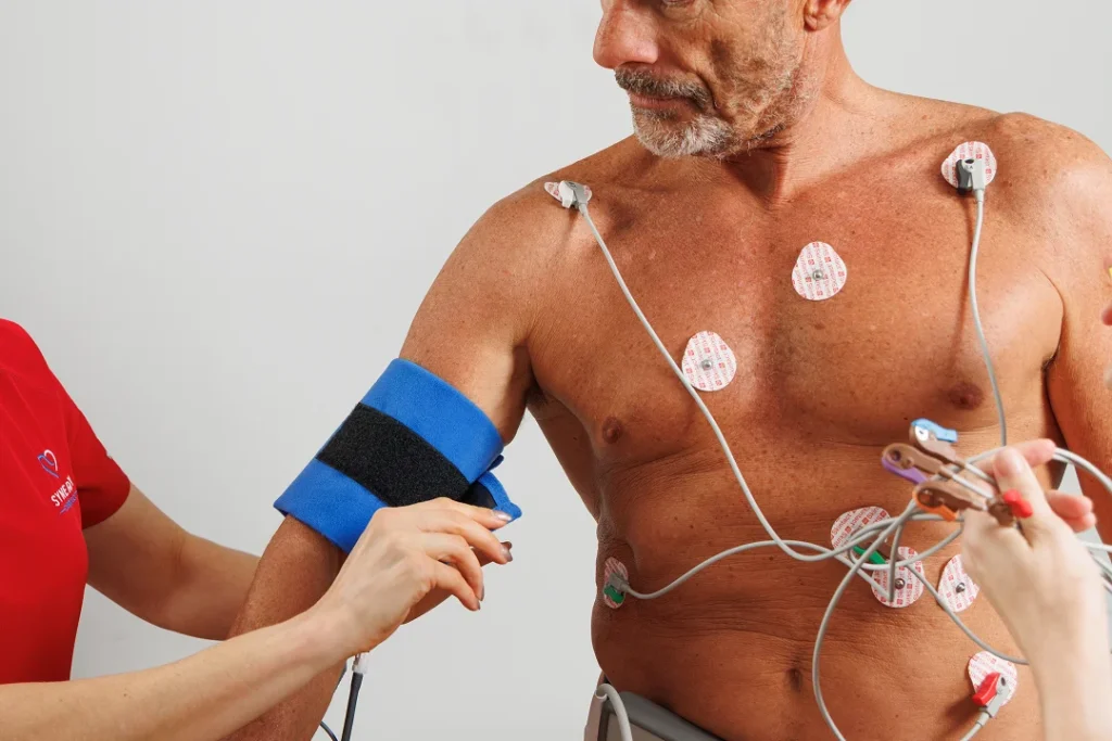
Baseline heart imaging is performed before exercise
Electrodes are placed on your chest to monitor your heart rhythm
You will walk on a treadmill while your heart rate, blood pressure and symptoms are monitored
Further heart images are taken immediately after exercise
The test is supervised by an experienced clinical team in a controlled environment.
Your cardiologist will review the results and provide a report to your referring doctor.
You may be referred for a stress echocardiogram if you have:
Chest pain or discomfort
Suspected coronary artery disease
Shortness of breath during exertion
Reduced exercise tolerance
Symptoms that occur during physical activity
It may also be recommended to assess heart function under stress or to investigate abnormal findings from other cardiac tests.
Dr Jodi Harker provides expert assessment of stress echocardiogram results to support accurate diagnosis and treatment planning.
Her focus is on clear interpretation, patient safety, and practical recommendations based on each individual’s symptoms and risk profile.
Please bring:
This helps ensure your cardiologist has all the information needed to provide accurate assessment and advice.
If you have been referred for a stress echocardiogram, our team is here to provide safe, structured and professional cardiac testing.
An Exercise Stress Echocardiogram, or stress echo, is a test that combines an ultrasound of your heart with a treadmill exercise test. It helps us see how well your heart functions when it’s working harder—such as during physical activity. The test shows how your heart muscles and valves respond under stress and helps detect any areas with poor blood flow.
Yes, the test is generally safe and is supervised by trained medical professionals. Your heart rhythm, blood pressure, and physical responses will be closely monitored throughout the procedure. Like any test involving exercise, there may be mild risks, especially if you have existing heart issues, but emergency care is readily available if needed.
Your doctor may recommend this test to evaluate symptoms like chest pain, shortness of breath, or fatigue during physical activity. It also helps us detect coronary artery disease, assess how well previous treatments like stents or bypass surgery are working, and determine your heart’s overall fitness and function.
Once the test is complete, you’ll be allowed to rest and recover. The cardiologist will review the images and measurements, then schedule a time to discuss the results with you. Based on the findings, further testing or treatment may be recommended if necessary.
Wear comfortable, loose-fitting clothes and walking or running shoes. Avoid wearing lotions or oils on your chest area on the day of the test, as these can interfere with the ultrasound imaging. It’s also helpful to bring a list of your current medications.
You may be asked not to eat or drink anything except water for a few hours before the test. Some medications, particularly those affecting your heart rate or blood pressure, may need to be paused. Your doctor or a nurse from Synergy Cardiology will provide clear instructions based on your health profile.